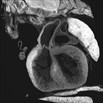
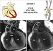
 | 2D serial EFIC image stack in the coronal view of mutant 449-005-2 (ED16.5) reveals PTA type-I
Click thumbnail to play movie. | Tab1b2b449Clo/Tab1b2b449Clo | C57BL/6J-Tab1b2b449Clo |
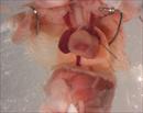 | Mutant 449-005-2 (E16.5) exhibit persistent truncus arteriosus (PTA), which was confirmed by EFIC analysis. | Tab1b2b449Clo/Tab1b2b449Clo | C57BL/6J-Tab1b2b449Clo |
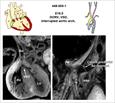
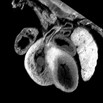 | 2D serial EFIC image stack in the coronal view of mutant 449-005-1 (ED16.5) reveals DORV {SDD}, subaortic VSD, and interrupted aortic arch type-B
Click thumbnail to play movie. | Tab1b2b449Clo/Tab1b2b449Clo | C57BL/6J-Tab1b2b449Clo |
 | Mutant 449-005-1 (E16.5) exhibit interrupted aortic arch (IAA) and malalignment of the great arteries, which was shown to be double outlet right ventricle (DORV) by EFIC analysis. | Tab1b2b449Clo/Tab1b2b449Clo | C57BL/6J-Tab1b2b449Clo |
 | | Tab1b2b449Clo/Tab1b2b449Clo | C57BL/6J-Tab1b2b449Clo |
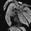 | 2D serial EFIC image stack in the coronal plane of newborn mutant 449-002-NA reveals DORV {SDD}, suboaortic VSD, Interrupted aortic arch type-B, and high take-off right coronary artery
Click thumbnail to play movie. | Tab1b2b449Clo/Tab1b2b449Clo | C57BL/6J-Tab1b2b449Clo |
 | Newborn mutant 449-002-NA exhibit interrupted aortic arch (IAA) and malalignment of the great arteries, confirmed as double outlet right ventricle (DORV) by EFIC analysis. | Tab1b2b449Clo/Tab1b2b449Clo | C57BL/6J-Tab1b2b449Clo |
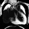 | 2D serial EFIC image stack in the coronal plane of newborn mutant 449-003-NB (ED21.5) reveals DORV {SDD}, subaortic VSD, ASD (II), and vascular sling.
Click thumbnail to play movie. | Tab1b2b449Clo/Tab1b2b449Clo | C57BL/6J-Tab1b2b449Clo |
 | | Tab1b2b449Clo/Tab1b2b449Clo | C57BL/6J-Tab1b2b449Clo |
 | | Tab1b2b449Clo/Tab1b2b449Clo | C57BL/6J-Tab1b2b449Clo |
 | | Tab1b2b449Clo/Tab1b2b449Clo | C57BL/6J-Tab1b2b449Clo |
 | 2D imaging of mutant 449-006-6 (E17.5) in transverse view showed atrioventricular septal defect (AVSD)
Click thumbnail to play movie. | Tab1b2b449Clo/Tab1b2b449Clo | C57BL/6J-Tab1b2b449Clo |
 | Color flow imaging of mutant 449-006-6 (E17.5) in transverse view showed infow tract regurgitation at endocardial cushion level suggesting atrioventricular septal defect (AVSD). | Tab1b2b449Clo/Tab1b2b449Clo | C57BL/6J-Tab1b2b449Clo |
 | Color flow imaging of mutant 449-006-6 (E17.5) in transverse view showed infow tract forward flow and regurgitation at endocardial cushion level suggesting atrioventricular septal defect (AVSD).
Click thumbnail to play movie. | Tab1b2b449Clo/Tab1b2b449Clo | C57BL/6J-Tab1b2b449Clo |
 | Color flow imaging of mutant 449-006-6 in transverse view showed infow tract forward flow at endocardial cushion level which indicates AVSD | Tab1b2b449Clo/Tab1b2b449Clo | C57BL/6J-Tab1b2b449Clo |
 | Spectral Doppler of mutant 449-006-6 (E17.5) showed inflow tract regurgitation (R). | Tab1b2b449Clo/Tab1b2b449Clo | C57BL/6J-Tab1b2b449Clo |
 | Color flow imaging of mutant 449-006-6 (E17.5) in sagittal view showed single outflow originating from the right ventricle (RV) with regurgitation, and a large VSD indicating persistent truncus arteriousus (PTA).
Click thumbnail to play movie. | Tab1b2b449Clo/Tab1b2b449Clo | C57BL/6J-Tab1b2b449Clo |
 | Color flow imaging of mutant 449-006-6 (E17.5) in sagittal view showed single outflow originating from the right ventricle (RV) and a large VSD indicating persistent truncus arteriosus (PTA). | Tab1b2b449Clo/Tab1b2b449Clo | C57BL/6J-Tab1b2b449Clo |
 | Color flow imaging of mutant 449-006-6 (E17.5) in sagittal view showed single outflow originating from the right ventricle with regurgitation, a large VSD, and persistent truncus arteriosus (PTA). | Tab1b2b449Clo/Tab1b2b449Clo | C57BL/6J-Tab1b2b449Clo |
 | Mutant 449-006-6 (E17.5) with interrupted aortic arch (IAA) and persistent truncus arteriosus (Van Praagh type A4) confirmed by EFIC analysis. | Tab1b2b449Clo/Tab1b2b449Clo | C57BL/6J-Tab1b2b449Clo |
 | | Tab1b2b449Clo/Tab1b2b449Clo | C57BL/6J-Tab1b2b449Clo |
 Analysis Tools


