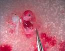
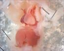
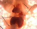
 | Mutant 1554-004-1 (E14.5) exhibits gastroschisis with liver and gut outside the abdominal cavity (celoschisis) | Lrp1b2b1554Clo/Lrp1b2b1554Clo | C57BL/6J-Lrp1b2b1554Clo |
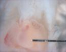
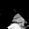
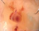 | Mutant fetus 1554-004-1 (E14.5) exhibits narrowed pulmonary outflow | Lrp1b2b1554Clo/Lrp1b2b1554Clo | C57BL/6J-Lrp1b2b1554Clo |
 | Mutant fetus 1554-004-1 (E14.5) exhibits narrowed pulmonary outflow | Lrp1b2b1554Clo/Lrp1b2b1554Clo | C57BL/6J-Lrp1b2b1554Clo |
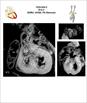
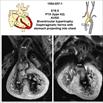
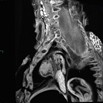
 | Serial 2D EFIC image stack in the coronal plane of 1554-004-1 (E14.5) reveals AVSD (or closing interventricular foramen), small PA
Click thumbnail to play movie. | Lrp1b2b1554Clo/Lrp1b2b1554Clo | C57BL/6J-Lrp1b2b1554Clo |
 | Mutant fetus 1554-004-1 (E14.5) exhibits gastroschsis with liver protruding outside of abdominal cavity (celoschisis) | Lrp1b2b1554Clo/Lrp1b2b1554Clo | C57BL/6J-Lrp1b2b1554Clo |
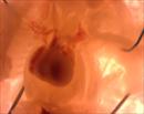
 | Mutant fetus 1554-4-2 exhibits narrowed pulmonary stenosis | Lrp1b2b1554Clo/Lrp1b2b1554Clo | C57BL/6J-Lrp1b2b1554Clo |
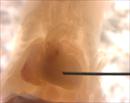 | Mutant 1554-004-2 (E14.5) shows pulmonary stenosis | Lrp1b2b1554Clo/Lrp1b2b1554Clo | C57BL/6J-Lrp1b2b1554Clo |
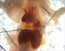
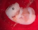 | Mutant fetus 1554-004-1 (E14.5) exhibits body wall closure defect with liver protruding outside of abdominal cavity (celoschisis) | Lrp1b2b1554Clo/Lrp1b2b1554Clo | C57BL/6J-Lrp1b2b1554Clo |
 | Mutant fetus 1554-004-1 (E14.5) exhibits body wall closure defect with liver protruding outside of abdominal cavity (celoschisis) | Lrp1b2b1554Clo/Lrp1b2b1554Clo | C57BL/6J-Lrp1b2b1554Clo |
 | EFIC Summary | Lrp1b2b1554Clo/Lrp1b2b1554Clo | C57BL/6J-Lrp1b2b1554Clo |
 | Serial 2D EFIC image stack of mutant fetus 1554-004-2 (E14.5) shows DORV with pulmonary stenosis and AVSD
Click thumbnail to play movie. | Lrp1b2b1554Clo/Lrp1b2b1554Clo | C57BL/6J-Lrp1b2b1554Clo |
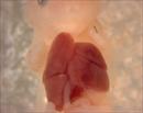
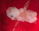
 | Mutant fetus 1554-007-1 (E14.5) exhibits single outflow tract with abdominal organ projecting into thoracic cavity, indicating a diaphragmatic hernia | Lrp1b2b1554Clo/Lrp1b2b1554Clo | C57BL/6J-Lrp1b2b1554Clo |
 | Mutant fetus 1554-007-1 (E14.5) exhibits single outflow tract with abdominal organ projecting into thoracic cavity, indicating a diaphragmatic hernia | Lrp1b2b1554Clo/Lrp1b2b1554Clo | C57BL/6J-Lrp1b2b1554Clo |
 | Mutant fetus 1554-007-1 (E14.5) exhibits single outflow tract with abdominal organ protruding into chest cavity indicating diaphragmatic hernia | Lrp1b2b1554Clo/Lrp1b2b1554Clo | C57BL/6J-Lrp1b2b1554Clo |
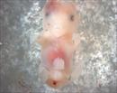 | Mutant 1554-007-1 exhibits umbilical herniation indicative of an omphalocele type body wall closure defect | Lrp1b2b1554Clo/Lrp1b2b1554Clo | C57BL/6J-Lrp1b2b1554Clo |
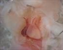 | Mutant fetus 1554-007-1 (E14.5) exhibits a single outflow tract, indicating persistent truncus arteriosus (PTA) | Lrp1b2b1554Clo/Lrp1b2b1554Clo | C57BL/6J-Lrp1b2b1554Clo |
 | EFIC Summary | Lrp1b2b1554Clo/Lrp1b2b1554Clo | C57BL/6J-Lrp1b2b1554Clo |
 | Serial 2D EFIC image stack in the coronal plane of 1554-007-1 (E16.5) reveals persistence truncus arteriosis (PTA/Type II) hypoplastic PA {S,D,D}
Click thumbnail to play movie. | Lrp1b2b1554Clo/Lrp1b2b1554Clo | C57BL/6J-Lrp1b2b1554Clo |
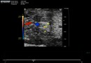 | Color flow spectral Doppler imaging of mutant 1554-007-1 (E16.5) in the frontal view shows a single outflow tract suggesting PTA with regurgitation and VSD
Click thumbnail to play movie. | Lrp1b2b1554Clo/Lrp1b2b1554Clo | C57BL/6J-Lrp1b2b1554Clo |
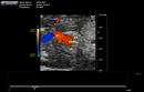 | Snapshot of color flwo spectral Doppler movie of mutant 1554-007-1 (E16.5) in the frontal view shows a single outflow tract suggesting PTA and VSD | Lrp1b2b1554Clo/Lrp1b2b1554Clo | C57BL/6J-Lrp1b2b1554Clo |
 Analysis Tools