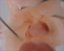
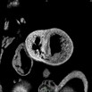
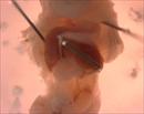
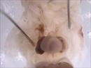
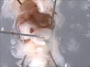
 | Mutant 386-005-1 presents with an anterior aorta which is diagnosed as DORV by EFIC imaging | b2b386Clo/b2b386Clo | C57BL/6J-b2b386Clo |
| b2b386.1Clo/b2b386.1Clo | C57BL/6J-b2b386.1Clo |
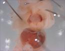 | Mutant 386-005-1 exhibits heterotaxy including left-sided IVC, right liver isomerism (1R/1L), and left lung isomerism (2R/2L) | b2b386Clo/b2b386Clo | C57BL/6J-b2b386Clo |
| b2b386.1Clo/b2b386.1Clo | C57BL/6J-b2b386.1Clo |
 | EFIC Summary | b2b386Clo/b2b386Clo | C57BL/6J-b2b386Clo |
| b2b386.1Clo/b2b386.1Clo | C57BL/6J-b2b386.1Clo |
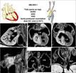
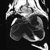
 | Serial 2D EFIC image stack in the coronal plane of 386-005-1 (E15.5) reveals DORV {A,D,D}, AVSD, and bilateral IVC
Click thumbnail to play movie. | b2b386Clo/b2b386Clo | C57BL/6J-b2b386Clo |
| b2b386.1Clo/b2b386.1Clo | C57BL/6J-b2b386.1Clo |
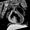
 | Serial 2D EFIC image stack in the transverse plane of 386-005-1 reveals DORV {A,D,D}, AVSD, and bilateral IVC
Click thumbnail to play movie. | b2b386Clo/b2b386Clo | C57BL/6J-b2b386Clo |
| b2b386.1Clo/b2b386.1Clo | C57BL/6J-b2b386.1Clo |
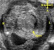
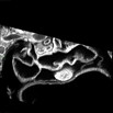
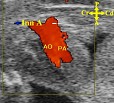 | Ultrasound imaging of mutant 386-005-1 reveals anterior aorta and posterior pulmonary artery | b2b386Clo/b2b386Clo | C57BL/6J-b2b386Clo |
| b2b386.1Clo/b2b386.1Clo | C57BL/6J-b2b386.1Clo |
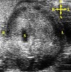
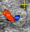 | Ultrasound imaging of mutant 386-005-1 reveals that the aorta originates from the right ventricle and a VSD | b2b386Clo/b2b386Clo | C57BL/6J-b2b386Clo |
| b2b386.1Clo/b2b386.1Clo | C57BL/6J-b2b386.1Clo |
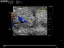 | Ultrasound imaging of mutant 386-005-1 reveals aorta originates from right ventricle with aortic regurgitation and the presence of a VSD
Click thumbnail to play movie. | b2b386Clo/b2b386Clo | C57BL/6J-b2b386Clo |
| b2b386.1Clo/b2b386.1Clo | C57BL/6J-b2b386.1Clo |
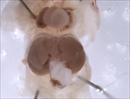
 | Ultrasound imaging of mutant 386-005-1 reveals heart apex points to the left, indicative of levocardia | b2b386Clo/b2b386Clo | C57BL/6J-b2b386Clo |
| b2b386.1Clo/b2b386.1Clo | C57BL/6J-b2b386.1Clo |
 | Ultrasound imaging reveals a right-sided stomach (S) | b2b386Clo/b2b386Clo | C57BL/6J-b2b386Clo |
| b2b386.1Clo/b2b386.1Clo | C57BL/6J-b2b386.1Clo |
 | Mutant 386-005-1 presents with dextrogastria and asplenia | b2b386Clo/b2b386Clo | C57BL/6J-b2b386Clo |
| b2b386.1Clo/b2b386.1Clo | C57BL/6J-b2b386.1Clo |
 | Mutant 386-005-1 presents with left liver isomerism | b2b386Clo/b2b386Clo | C57BL/6J-b2b386Clo |
| b2b386.1Clo/b2b386.1Clo | C57BL/6J-b2b386.1Clo |
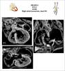
 | Mutant 386-005-2 (E15.5) exhibits heterotaxy with left lung isomerism (1R/1L) and dual IVC | b2b386Clo/b2b386Clo | C57BL/6J-b2b386Clo |
| b2b386.1Clo/b2b386.1Clo | C57BL/6J-b2b386.1Clo |
 | EFIC Summary | b2b386Clo/b2b386Clo | C57BL/6J-b2b386Clo |
| b2b386.1Clo/b2b386.1Clo | C57BL/6J-b2b386.1Clo |
 | Serial 2D EFIC image stack in the coronal plane of 386-005-2 (E15.5) reveals AVSD, common atrium, and bilateral IVC
Click thumbnail to play movie. | b2b386Clo/b2b386Clo | C57BL/6J-b2b386Clo |
| b2b386.1Clo/b2b386.1Clo | C57BL/6J-b2b386.1Clo |
 | Mutant 386-005-2 (E15.5) exhibits left liver isomerism (2R/2L) | b2b386Clo/b2b386Clo | C57BL/6J-b2b386Clo |
| b2b386.1Clo/b2b386.1Clo | C57BL/6J-b2b386.1Clo |
 | Mutant 386-005-2 (E15.5) exhibits asplenia | b2b386Clo/b2b386Clo | C57BL/6J-b2b386Clo |
| b2b386.1Clo/b2b386.1Clo | C57BL/6J-b2b386.1Clo |
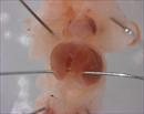
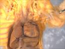 | Mutant 386-007-ND shows an enlarged right atrium and abnormal arrangement of the outflow tracts which is diagnosed as DORV by EFIC imaging | b2b386Clo/b2b386Clo | C57BL/6J-b2b386Clo |
| b2b386.1Clo/b2b386.1Clo | C57BL/6J-b2b386.1Clo |
 | EFIC Summary | b2b386Clo/b2b386Clo | C57BL/6J-b2b386Clo |
| b2b386.1Clo/b2b386.1Clo | C57BL/6J-b2b386.1Clo |
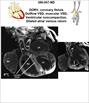
 | Serial 2D EFIC image stack in the coronal plane of 386-007-ND reveals DORV, coronary fistula, outflow VSD, muscular VSD, ventricular myocardial non-compaction, and dilated atria/venous return
Click thumbnail to play movie. | b2b386Clo/b2b386Clo | C57BL/6J-b2b386Clo |
| b2b386.1Clo/b2b386.1Clo | C57BL/6J-b2b386.1Clo |
 Analysis Tools